
We turn tissue images into quantitative assessments.
Aikov Labs builds AI tools that automate time-consuming tissue analysis for pathology laboratories and research teams. The pathologist reviews every result.
The estimate becomes a measurement.
Our models segment the tissue, detect each cell and classify it by its staining. On this Ki-67 image, Cell Analytics classified 87,844 nuclei; the positive ones are shown in magenta.
A proportion that is often judged by eye becomes a number the pathologist can check, correct and repeat.
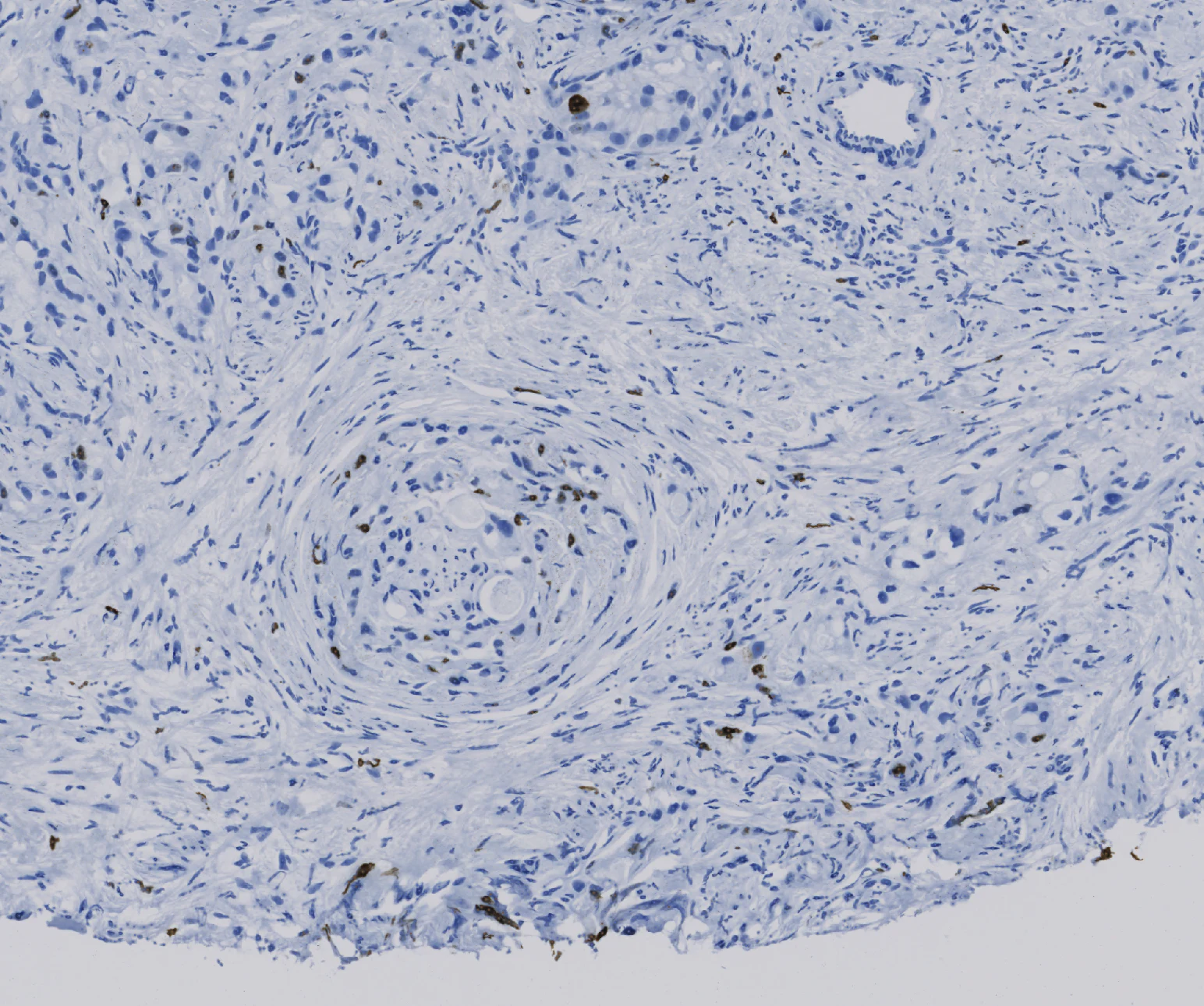

Cell Analytics result, this image
| Ki-67 positive | 2,274 | 2.6% |
|---|---|---|
| Ki-67 negative | 85,570 | 97.4% |
Two tools, one method.
-
Cell Analytics
Tissue segmentation, cell detection and quantitative counting in H&E and immunohistochemistry images, for biomarkers such as Ki-67.
-
TIL Analytics
Quantification of tumor-infiltrating lymphocytes, starting with triple-negative breast cancer, with spatial maps of tumor, stroma and lymphocytes for review.
Working with digital slides? We would like to hear from you.
We work with pathology laboratories, hospital departments and research groups on focused evaluations of our tools.
info@aikovlabs.com